


 61 / 176

705
ANATOMICAL VARIATIONS OF CENTRAL VEINS
Congenital
The commonest SVC variant is a left sided vein, which occurs
with or without a normal right SVC (0.5% population, and
higher with cardiac defects). A left SVC crosses the arch of the
aorta and left pulmonary hilum, and enters the right atrium
FIGURE 5B. INSET CT IMAGE.
Catheter was pulled back 3cm but was still seen to be abutting side wall
of innominate vein.
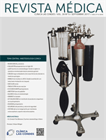
FIGURE 6. AXIAL CT OF CHEST SHOWING THE LEFT
INNOMINATE VEIN CURVING ANTERIORLY TO ADD
FURTHER CORNERS FOR LEFT SIDED CATHETER TO
PASS THROUGH TO ACCESS THE SVC
This patient had distorted aortic arch from aneurysm but this curvature
is seen without obvious disease. It is a common reason for difficulty in
positioning left sided catheters.
via an enlarged coronary sinus. It can be used for access if
entering right atrium, but may open into the left atrium with
risks of systemic embolism (17). The IVC can show similar
double variation.
Patients with dextrocardia have the heart orientated in
reverse so that it lies over to the right. It can be associ-
ated with the reversal of abdominal/chest organs and blood
vessels, so-called situs inversus, in which case the SVC and
IVC also lie to the left.
Acquired
Acute SVC compression from tumour can cause oedema and
venous engorgement in the upper body (SVC syndrome).
Stenosis or thrombosis is common with long-term access,
and is often asymptomatic due to collateral vein forma-
tion. This can present as a failure to pass a guidewire or
catheter.
Obvious venous collaterals on the chest wall, difficult to
compress veins on ultrasound, or higth venous pressure on
cannulation suggest this problem. Confirmation is by venog-
raphy, CT, or Doppler ultrasound studies (Figure 7). Medias-
tinal shift from effusions, lung collapse, or pneumonectomy
will shift mediastinal structures including the SVC. In the
event of IV blockage, the azygous system enlarges to provide
drainage.
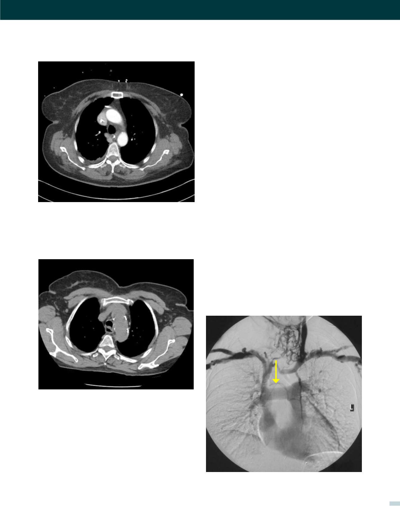
There in blockage of the left innominate vein (arrow) with collateral flow
around the thyroid area and within the chest.
FIGURE 7. A PATIENT HAVING CONTRAST INJECTED UO
BOTH ARMS SIMULTANEOUSLY
[VASCULAR ACCESS - DR. ANDREW BODENHAM]