


 60 / 176

704
UPPER ARM VEINS
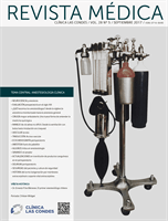
High resolution ultrasound easily identifies deeper veins in
the proximal upper arm to aid access in difficult case, and
PICC insertion in the upper arm to avoid elbow flexure PICCs
can be inserted from antecubital fossa with direct vision. The
basilic, brachial, and cephalic veins are seen with arteries and
nerves (Figure 3). Note close relation of median nerve and
brachial veins, and cutaneous nerve of forearm and basilic
vein. The cephalic vein runs a tortuous course to enter the
axillary vein leading to difficulties in passing catheters. There
is wide anatomical variation.
the vein wall and cause a hydrothorax (Figure 5). The lower
section of the SVC is within the pericardium so a perforation
risks cardiac tamponade.
The azygous vein ascends on the right side in the
posterior mediastinum, passes anteriorly to join the
mid-section of SVC above the hilum and is a site for tip
malposition. Left sided catheters traverse one or more
corners to pass to the SVC, making tip placement more
difficult, particularly if the left innominate vein curves
anteriorly (Figure 6).
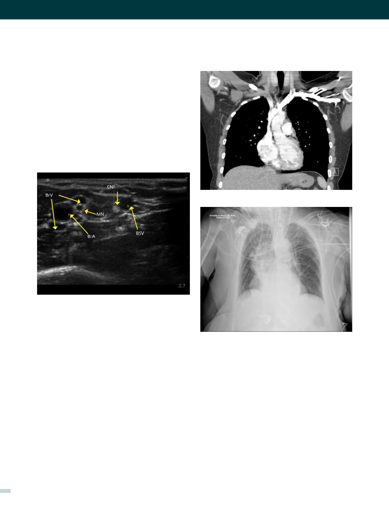
FIGURE 4. CORONAL CT OF CHEST SHOWING CLOSE
PROXIMITY OF SVC TO PLEURA AND ASCENDING AORTA
FIGURE 5A.
Access is easiest with a sharp small-bore needle (20g), fine
guidewire, dilator, and sheath (micropuncture set). The cath-
eter is measured for length externally, from X-ray screening
or ECG guidance (15).
Applied anatomy of the superior vena cava (SVC)
The lower SVC is the target for catheter tips from the upper-
body and applied anatomy is important (16). It is formed by
the two brachiocephalic veins behind the first right costal
cartilage. It is approximately 2cm in diameter and 7cm long
with no valves and descends to the right atrium (Figure 4). Its
right border is partially visible on chest X-ray but it is difficult
to visualize the junction with right atrium.
The upper right border of the SVC bulges into the low
pressure pleural space so a tear can cause major bleeding.
Abutting catheter tips (particularly from the left) can erode
a. A dialysis catheter
has been inserted from the left IJV and is too short
with catheter abuttting SVC wall, with risks of perforation and thrombosis.
The patient has large hiatus hernia.
[REV. MED. CLIN. CONDES - 2017; 28(5) 701-712]
FIGURE 3. US IMAGES OF RIGHT UPPER ARM VESSELS
UPPER ARM SEEN FROM BELOW
Brachial artery (BrA), Brachial veins (BrV), Basilic vein (BsV), Median nerve
MN, cutaneous nerves forearm (CNF).